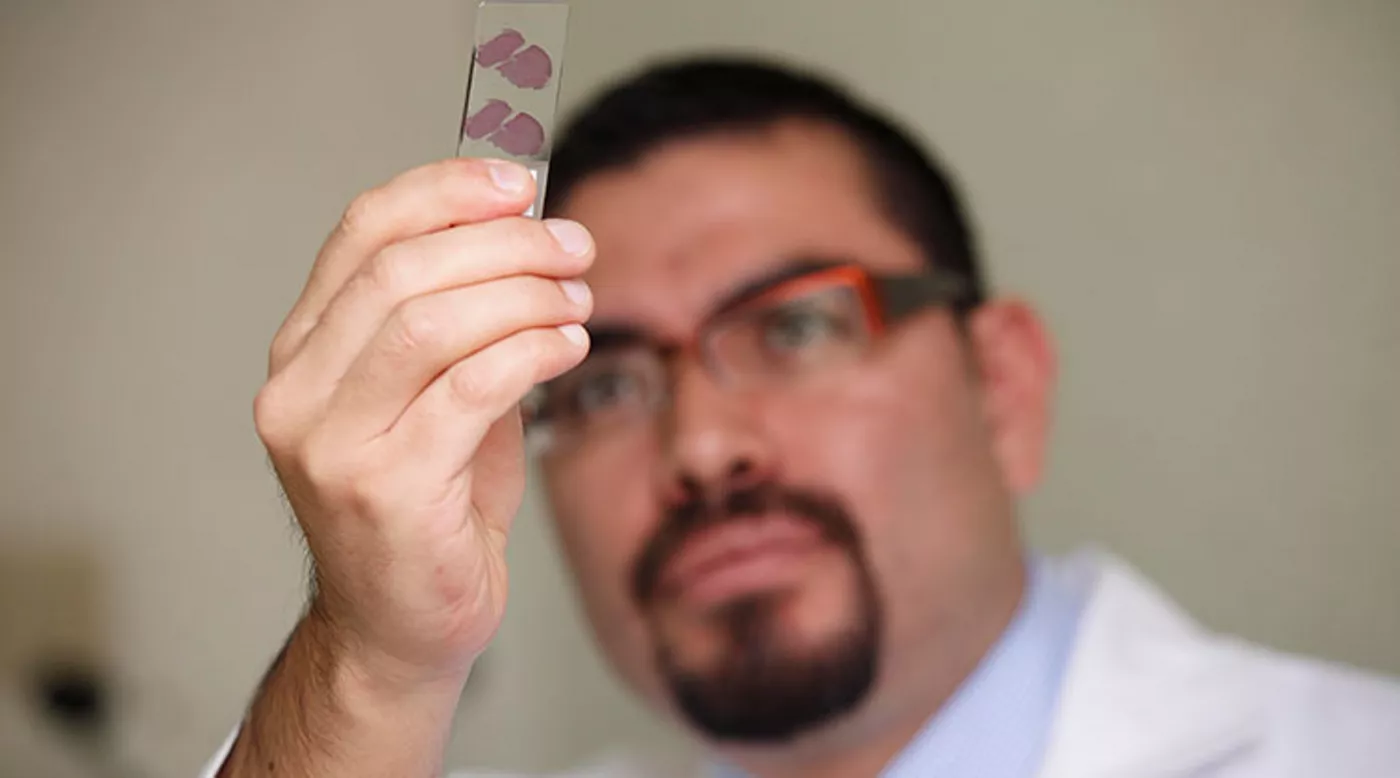
Pemphigus Vulgaris (PV): Pathology & Mechanisms of Disease
Anatomy of skin and oral mucosa
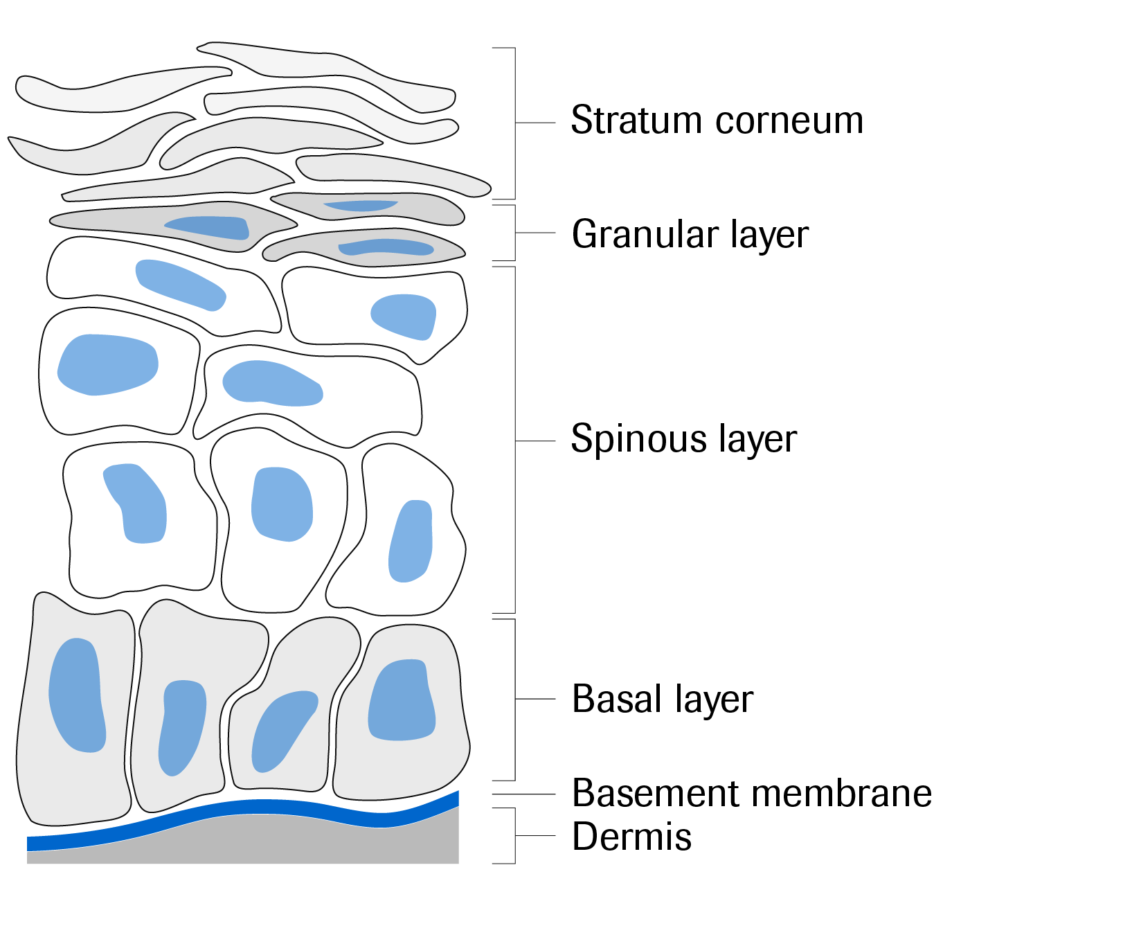
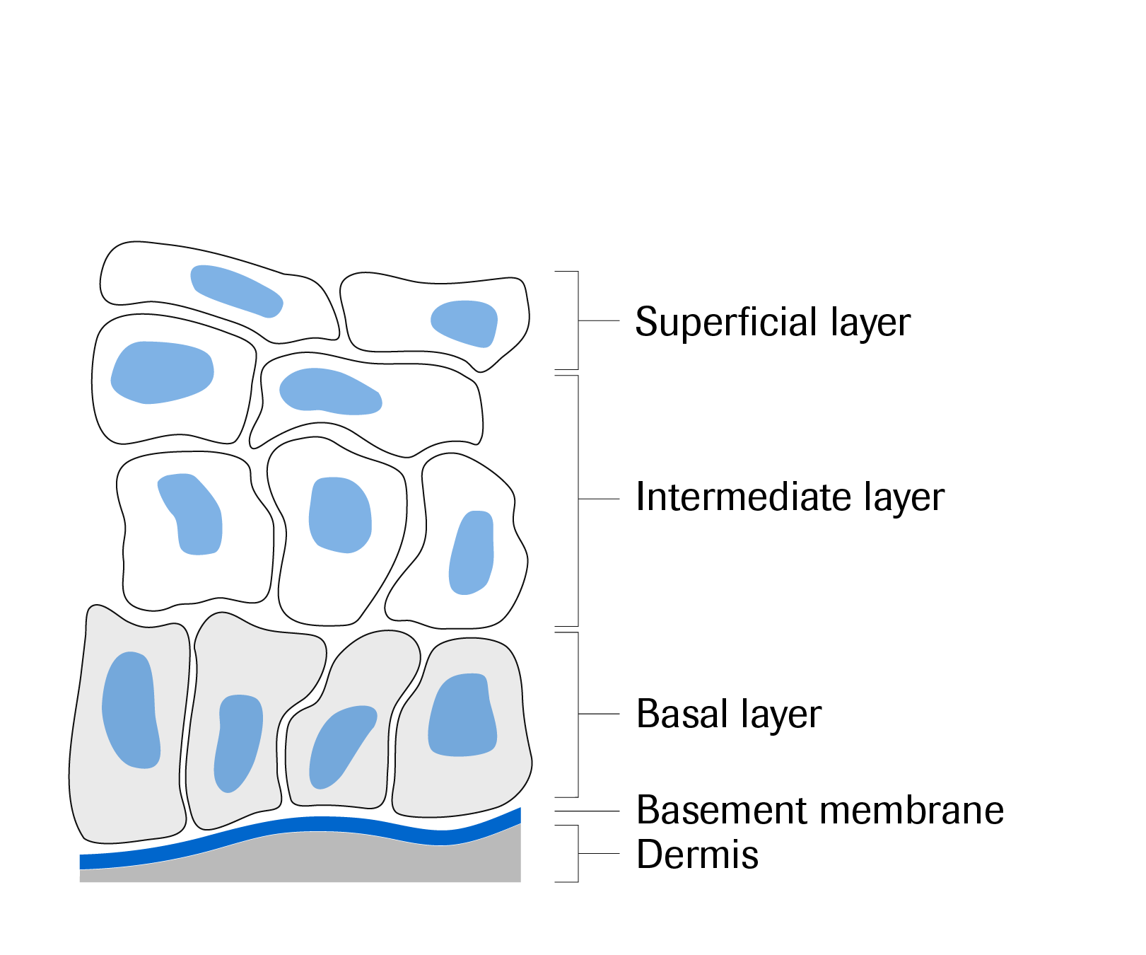
The epithelia of the skin and oral mucosa are comprised of multiple layers of cells. A key difference between the skin and oral mucosa is that the skin has a thick layer of dead keratinocytes, the stratum corneum, that functions as a protective barrier against pathogens and dehydration.1
Skin
Buccal mucosa
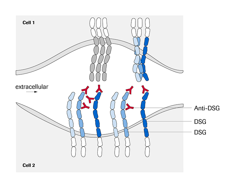
In the epidermis of normal healthy skin and mucosal membranes, cells are bound to each other by desmosomes and to the extracellular matrix by hemidesmosomes. Cell-cell adhesion in the desmosomes is facilitated by desmoglein proteins that form bonds between neighboring epithelial cells.2
Normal desmosome
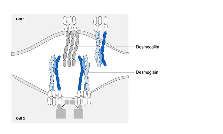
Figure adapted with permission from Kasperkiewicz M, et al. Nat Rev Dis Primers. 2017;3:17026.
Autoimmune cause of PV
PV is an autoimmune disease and caused by the development of immunoglobulin G (IgG) antibodies against the extracellular domains of desmogleins, specifically desmoglein 1 (DSG1) and desmoglein 3 (DSG3).3 B cells play a significant pathophysiological role in PV, as antibodies are produced by B cells as part of the adaptive immune response, including the production of autoantibodies in PV (anti-DSG3 and anti-DSG1)3. The autoantibodies to DSG1 and DSG3 disrupt the DSG-mediated binding between epithelial cells driving the loss of cell-cell adhesions between keratinocytes, a process known as acantholysis. It is as a result of acantholysis that severe blisters develop on the skin and/or mucosal membranes.3
Anti-DSG antibodies are hypothesized to result in acantholysis through two mechanisms: (1) auto-antibody-mediated steric hindrance of desmosomal adhesion and/or interference with desmosome assembly, and (2) intracellular cell signaling pathways that augment the autoimmune response.3
Steric hindrance
Intracellular signaling
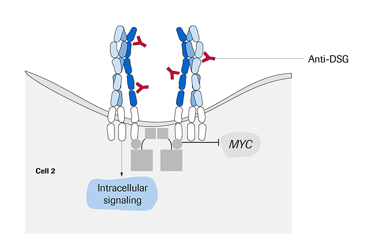
Figure adapted with permission from Kasperkiewicz M, et al. Nat Rev Dis Primers. 2017;3:17026.
DSG1 and DSG3 expression
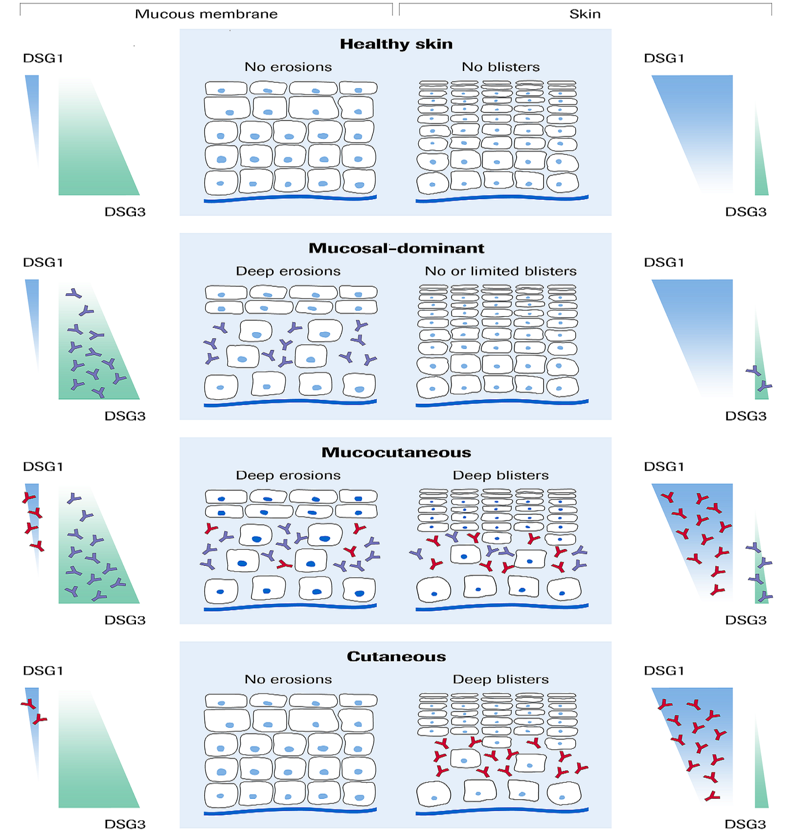
DSG1 and DSG3 are expressed differently in the skin and mucous membranes. DSG3 is expressed more highly than DSG1 in the mucous membranes. In the skin, DSG1 is expressed in the spinous and granular layers and is more abundant than DSG3, which is limited to the basal layers.3
In patients with PV, blisters arise in layers corresponding with the locations of DSG1 or DSG3 in the skin or mucosa. PV can be further subdivided into mucosal-dominant, mucocutaenous or cutaneous type.3 The cutaneous form is less common than the other forms which also involve the mucosal membranes.3
- Mucosal-dominant: Predominantly or only anti-DSG3 antibodies present affecting the mucosal membranes with no or limited skin involvement.
- Mucocutaneous: Anti-DSG1 and anti-DSG3 antibodies are present resulting in erosions to the mucosa and blistering of the skin.
- Cutaneous: Both anti-DSG1 and anti-DSG3 antibodies are present but the anti-DSG3 antibodies are pathogenically weak in the presence of potent anti-DSG1 antibodies, limiting involvement to the skin.
To learn more about the pathophysiology of PV, please watch the video
References
- MacNeal RJ. Available at: https://www.msdmanuals.com/en-gb/home/skin-disorders/biology-of-the-skin/structure-and-function-of-the-skin. Accessed December 2018.
- Green KJ, et al. Faseb j. 1996; 10:871-881.
- Kasperkiewicz M, et al. Nat Rev Dis Primers. 2017; 3:17026.
